
Thrombolysis and mechanical thrombectomy have revolutionized acute ischemic stroke management in the last two decades. And new AI-powered technologies such as RapidAI are helping physicians enhance stroke diagnosis and workflow.
Here’s a look back at some of the publications from this year by physicians using RapidAI, showing us how the software is making a difference in stroke care.
- Faster and accurate detection of stroke
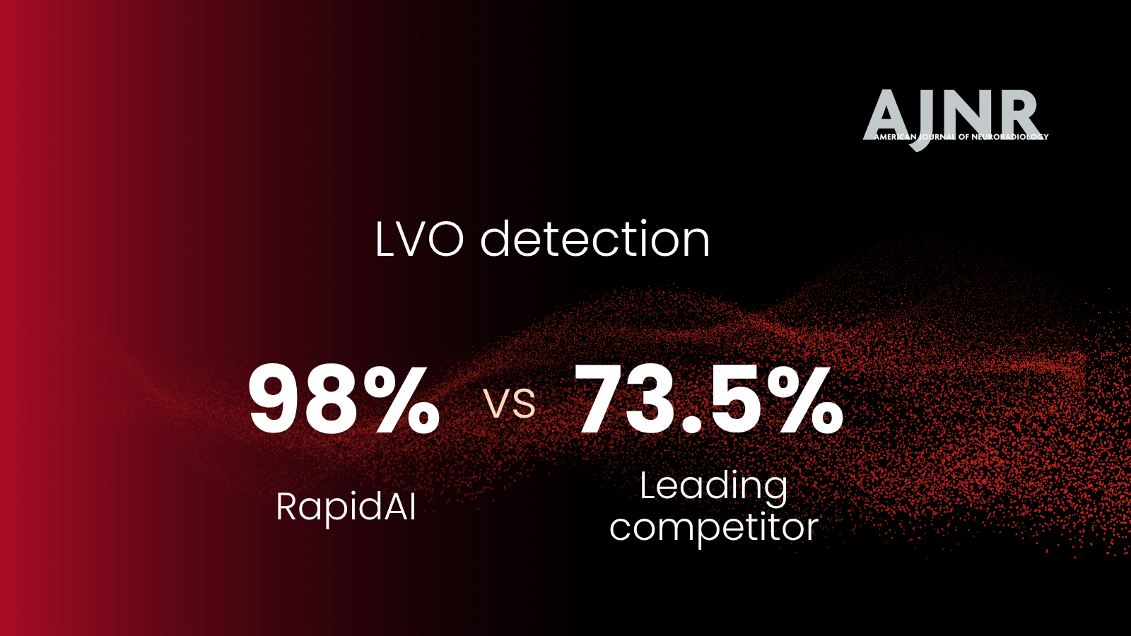
- Rapid LVO can quickly detect anterior circulation large vessel occlusions (LVOs) with high sensitivity and specificity
A retrospective study was carried out by Dr. Seena Dehkharghani and co-authors to investigate the ability of the automated software Rapid LVO to detect anterior circulation LVO on CTA scans from several institutions worlwide.
They found that Rapid LVO had a high sensitivity of 96% and specificity of 98% across subgroups – age, sex, imaging systems, and location. Rapid LVO quickly (mean average time was 3 minutes 30 seconds) detected anterior circulation LVOs without any user intervention.
- Providing Tmax maps using Rapid CTP software facilitates detection of distal vessel occlusions (DVOs) on CTA
Dr. Shalini Amukotuwa and co-authors carried out a retrospective study to determine whether using CTP-derived-Tmax maps improved diagnosis of DVOs.
The retrospective study showed that providing Tmax maps increased mean sensitivity for DVO detection from 70.7% to 90.4% and increased mean specificity from 87.5% to 95.7%. It also significantly improved diagnostic confidence and accuracy for the test readers.
- Rapid ICH accurately detects intracranial hemorrhage (ICH)
Due to the lack of immediate access to specialists who can review NCCT scans, ICH diagnosis can be missed, or may not be identified on time. Dr. Jeremy Heit, Dr. Francisco Mont’Alverne and co-authors conducted a retrospective study to evaluate whether the automated software Rapid ICH can help physicians by accurately detecting ICH and notifying them.
Rapid ICH had a high sensitivity of 96% and high specificity of 95%. The positive predictive value was 95.6% and negative predictive value was 95.3%. The positive likelihood ratio and negative likelihood ratio were favorable as well.
- Faster time to treatment
- Rapid mobile app significantly decreases intrahospital treatment times
Dr. Mais Al-Kawaz and co-authors investigated the impact of using Rapid clinical platform and mobile app on the intrahospital treatment times and stroke workflow at Johns Hopkins Hospital.
The Rapid mobile app significantly decreased door to groin puncture time by 33 minutes, door to first pass time by 35 minutes, and door to reperfusion time by 37 minutes. Following the deployment of Rapid mobile app, patients’ outcomes were also significantly improved
- With high positive predictive value (PPV) for LVO detection, Rapid CTA helps reduce treatment times and improves patient outcomes
Dr. Julie Adhya and co-authors evaluated their one-year experience using Rapid CTA and its impact on treatment times and patient outcomes at Allegheny Health Network, a comprehensive stroke center.
Rapid CTA demonstrated high PPV for LVO detection at a threshold of <45% relative vessel density, Using Rapid CTA decreased the CTA to groin puncture time significantly from 92 minutes to 68 minutes, thereby improving patient outcomes. Rapid CTA also helped facilitate worklist reprioritization.
- Aids patient selection for endovascular thrombectomy (EVT)
- Rapid for Angio could help select LVO patients eligible for EVT after direct transfer to angio suite
The potential of Rapid CTP to help select LVO patients eligible for endovascular thrombectomy has been validated in multiple clinical trials. A pilot study by Dr. Darko Quispe-Orozco and co-authors evaluated whether Rapid for Angio’s analysis of cone-beam CT perfusion (CBCTP) data were comparable to Rapid CTP’s analysis of multidetector CT perfusion (MDCTP) data.
The results showed that Rapid for Angio’s analysis of CBCTP data and Rapid CTP’s analysis of MDCTP data were comparable. There was a strong correlation between the ischemic core and hypoperfused tissue volumes. Rapid for Angio optimized for CBCTP could help facilitate stroke workflow and stroke care.
- Rapid MRI and Rapid CTP could help select basilar artery occlusion (BAO) patients eligible for EVT
A multicenter retrospective cohort study was performed by Dr. Carlo Cereda and co-authors to examine whether BAO patients with limited regions of severe hypoperfusion can benefit from EVT.
BAO patients with ≤ 3 Critical Area Perfusion Score (CAPS) and limited area of hypoperfusion (Tmax > 10 seconds) had favorable response to reperfusion following EVT. Reperfused CAPS ≤ 3 patients were more likely to achieve favorable functional outcomes at 90 days following EVT.
- Internationally adopted AI-powered stroke solution
- Successful implementation of Rapid software at a comprehensive stroke center in Buenos Aires, Argentina
Dr. Juan José Cirio and co-authors evaluated their experience with Rapid software in screening patients with hyperacute stroke at a comprehensive stroke center, Eneri Medical Institute at Buenos Aires, Argentina.
2,062 analyzes were performed. The software diagnosed 85 large vessel occlusions and 149 ICH. The mean ASPECT score was 8.4 (±2.1). 113 reperfusions were performed. RapidAI software presented the imaging data to physicians in a quick and secure manner, facilitating stroke diagnosis and treatment.
- Supports clinical research and advancements in stroke management
- The AURORA analysis shows late window mismatch patients have greater treatment effect from thrombectomy
Dr. Greg Albers and co-authors determined the optimal imaging profile to identify patients who would benefit from EVT in the late window (6-24 hours). Data was pooled from six randomized clinical trials and analyses were performed. All CTP and MRI scans of patients included in the AURORA database were processed using Rapid software.
Patients who met the DAWN (clinical mismatch) or DEFUSE 3 (Target mismatch) imaging profiles and were treated with EVT in the late window (6-24 hours) showed significant improvements in clinical outcomes. This treatment effect was significantly larger than observed in patients who did not have perfusion imaging and therefore had an undetermined imaging profile.
More patients fit the Target mismatch profile than the clinical mismatch profile and the results of this study show that Target mismatch can be used up to 24 hours to select patients.
- Real world data from Japan confirms the outcomes of mechanical thrombectomy up to 24 hours in LVOs
Dr. Manabu Inoue and co-authors retrospectively compared the clinical and radiological outcomes in early- (<6 hours) versus late- (6-24 hours) presenting LVO patients who underwent EVT. All diffusion weighted imaging and MRI/CTP images were processed using Rapid software.
The findings from a real‐world setting showed that both early and late time window patients selected for thrombectomy with automated perfusion imaging had clinical outcomes comparable with those from randomized trials, despite extending the window from 16 to 24 hours. The study also found that patients in the 6–24-hour time window had slower infarct growth rate.
- Rapid’s automated collateral measurements predict infarct growth rates
The study by Dr. Adam MacLellan and co-authors evaluated the association between baseline perfusion imaging collaterals (Hypoperfusion Intensity Ratio [HIR], Cerebral Blood Volume [CBV] index) and infarct growth at 24 hours in patients who did not achieve successful reperfusion in the DEFUSE 3 clinical trial. The association between baseline HIR and CBV index and clinical outcomes were also assessed.
The results showed that HIR threshold of 0.34 (AUC=0.68) and CBV index of 0.74 (AUC=0.72) can optimally predict infarct growth at 24 hours. Patients with favorable perfusion collaterals (HIR < 0.34 and CBV index > 0.74) had significantly less infarct growth at 24 hours. Baseline HIR and CBV index were not associated with 90-day functional outcome.
Assessing perfusion imaging collaterals using software such as Rapid can help predict evolution of stroke in the extended time window and guide patient selection.