
Radiology
Built for the realities of today’s radiologist


Designed around how radiologists actually read
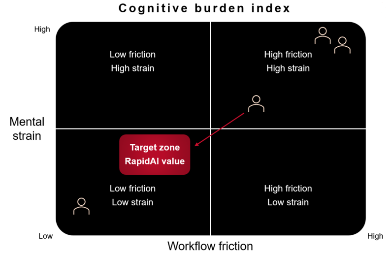
Navigator Pro gives radiologists a meaningful head start, showing where to look first, providing quantification up front, and reducing the strain of starting every case from scratch.
How it helps:
- Surfaces key findings and context early
- Cuts down the manual steps that drain energy
- Keeps prioritization, findings, communication, and reporting in one workspace
- Supports consistent, high-quality reads across long shifts

Broad disease state coverage, in a single workflow
Beyond our flagship solutions, we offer validated partner algorithms—seamlessly integrated into your workflow.
ORTHOPEDICS
Rapid VCF
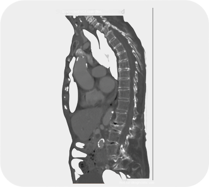
Detect suspected vertebral compression fractures in routine CT scans
- Address fractures early to improve outcomes and lower costs
Rapid C-Spine
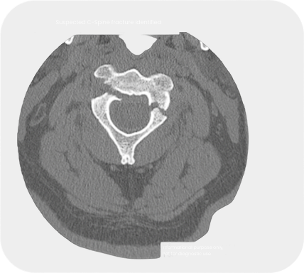
Identify suspected cervical spine fractures from NCCT imaging
- Reduce the delay between scan and interpretation for prompt medical attention
PULMONARY EMBOLISM
Rapid iPE
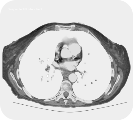
Review potential pulmonary embolisms in non-suspected cases
- Avoid delayed and missed findings, especially in cancer patients
Broad disease state coverage, in a single workflow
RapidAI delivers AI-driven insights across neurovascular, aortic, spine, chest, and cardiothoracic imaging—so radiologists can interpret a wide range of findings without switching systems or adding extra steps.
Neurovascular
Neurovascular
- Intracerebral hemorrhage (ICH)
- Subdural hematoma (SDH)
- Midline shift (MLS)
- Vessel occlusion (LMVO) / LVO/ MeVO
- NCCT LVO support
- Aneurysm
- DeltaFuse™ NCCT comparison
- Cranial fracture
- Obstructive hydrocephalus
Aortic
Aortic
- Aortic measurements
- Aortic morphology analysis

3D Reconstruction
3D Reconstruction
- Automated CTA reconstruction (head, neck, aortic arch)
- Curved planar reformats
- Vessel views + volume renderings
- Bone removal + segmentation

Spine
Spine
- C-spine fracture
- Vertebral compression fracture (VCF)

Chest & Cardiopulmonary
Chest & Cardiopulmonary
- Pulmonary embolism (PE)
- Incidental PE support (iPE)
- Lung nodules
- Nodule change analysis
- Pneumothorax
- Endotracheal tube (ETT) malposition
- Coronary artery calcium (CAC)
- Interstitial lung disease (ILD)
- COPD
- Emphysema
Broad disease state coverage, in a single workflow
Neurovascular
Aortic
3D Reconstruction
Spine
Chest & Cardiopulmonary
Musculoskeletal
Breast

Neurovascular
- Intracerebral hemorrhage (ICH)
- Subdural hematoma (SDH)
- Midline shift (MLS)
- Vessel occlusion (VO+) / LVO/ MeVO
- NCCT LVO support
- Aneurysm
- DeltaFuse™ NCCT comparison
- Cranial fracture
- Obstructive hydrocephalus
Aortic
- Aortic measurements
- Aortic morphology analysis
3D Reconstruction
- Automated CTA reconstruction (head, neck, aortic arch)
- Curved planar reformats
- Vessel views + volume renderings
- Bone removal + segmentation
Spine
- C-spine fracture
- Vertebral compression fracture (VCF)
Chest & Cardiopulmonary
- Pulmonary embolism (PE)
- Incidental PE support (iPE)
- Lung nodules
- Nodule change analysis
- Pneumothorax
- Endotracheal tube (ETT) malposition
- Coronary artery calcium (CAC)
- Interstitial lung disease (ILD)
- COPD
- Emphysema
Musculoskeletal
- Rib fracture
- Extremity fracture
Neurovascular
- Breast lesions
Trusted by radiologists worldwide