


A retrospective study showed that adding Tmax parameter and providing Tmax maps using Rapid CTP software facilitates detection of distal vessel occlusion (DVO) on CT angiography (CTA).
The findings by Dr. Shalini Amukotuwa and co-authors were published in Journal of Neuroradiology.
Key takeaways:
- Providing Tmax maps using Rapid CTP can facilitate detection of DVO on CTA
- With the addition of Tmax map, mean sensitivity for DVO detection went up from 70.7% to 90.4% and mean specificity went up from 87.5% to 95.7%.
- Diagnostic accuracy improved significantly for all four test readers with different levels of experience.
140 patients who had DVOs and whose CT, CTA, and CT perfusion (CTP) scans were available, were included in this study. Occlusions in the following arteries were considered as DVO: A2 to A5 segments of anterior cerebral artery; M2 to M4 segments of middle cerebral artery; P2 to P4 segments of posterior interior cerebral artery.
Two experienced neuroradiologists set up the reference standard. They were provided with patients’ clinical information and all available images.
Four test readers with different levels of experience independently reviewed the scans. The test readers included a second-year radiology resident, neuroradiology fellow, attending radiologist, and an imaging scientist.
They were provided with patients’ neurological deficits to mimic clinical setting.
When a Tmax map was available prior to CTA interpretation, the test readers were asked to localize the likely occluded segment based on the Tmax delay, and then carry out a focused search on CTA. If the Tmax map was not available prior to CTA interpretation, CTA was assessed for the presence (or absence) of DVO and its location reported. The test readers also rated diagnostic confidence following each CTA evaluation.
The test readers’ CTA analysis was compared against the reference standard.
Adding Tmax map improved sensitivity for DVO diagnosis
The gain in diagnostic sensitivity with Tmax map was even larger when only the data from the most distal occlusions were included in the analysis, and M2 and P2 were excluded. For instance, when proximal M2 occlusions were excluded, with the addition of Tmax map, mean sensitivity went up from 61% to 86.5%. When M2 and P2 occlusions were excluded, with the addition of Tmax map, diagnostic sensitivity went up from 42.6% to 81.5%.
Inter-reader agreement on CTA interpretation improved with the addition of Tmax map. Diagnostic accuracy and confidence improved significantly for all four test readers. Diagnostic speed improved significantly for the imaging scientist and neuroradiology fellow, the two test readers who were timed.
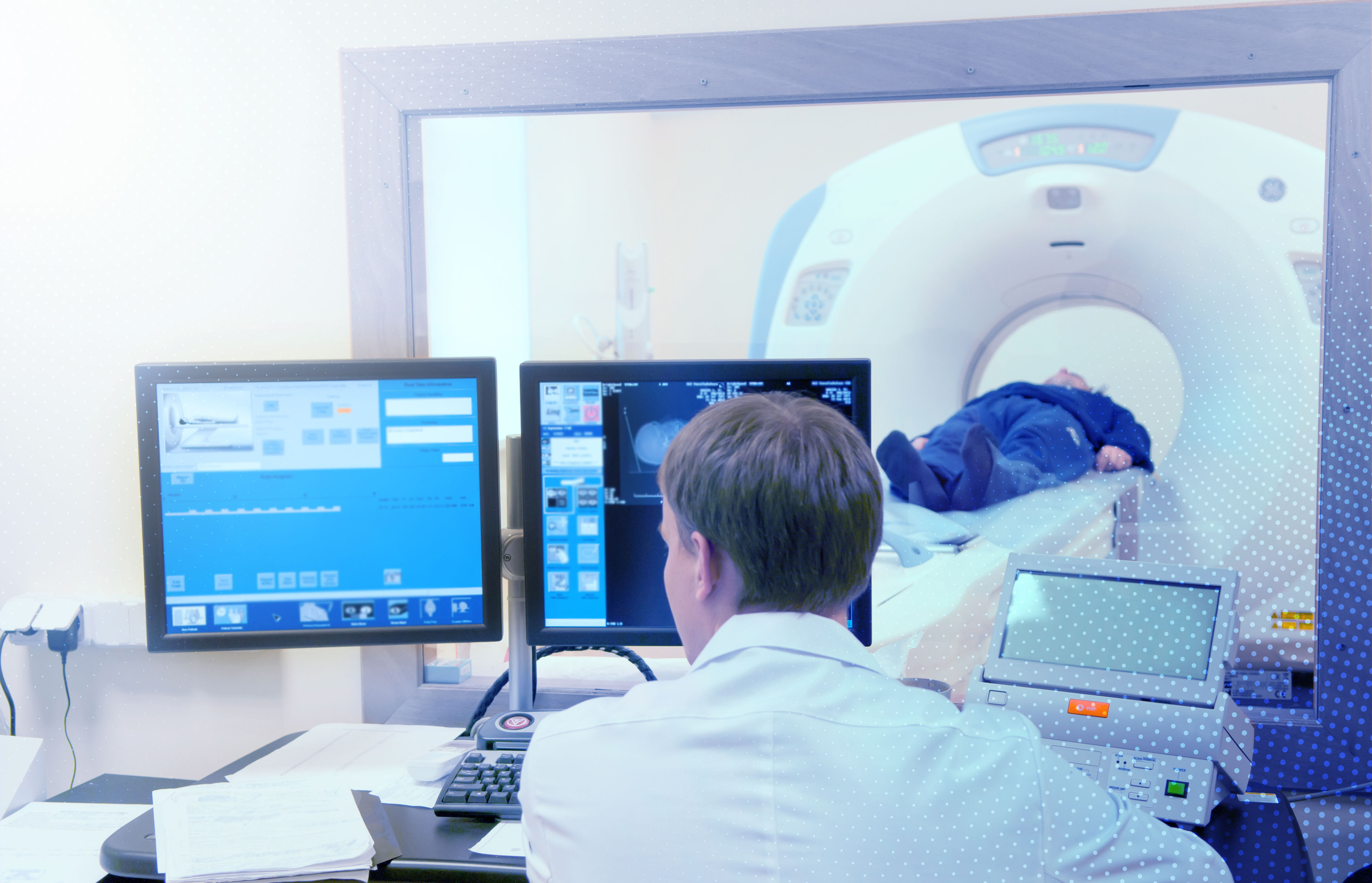
CTA has now become an essential component of acute stroke imaging protocol. However, detecting DVO on CTA can be challenging. With advances in endovascular device technology, it is now possible to treat obstructions in smaller, distal arteries in select patients. Quick detection of DVO is essential to perform a risk-benefit assessment and create a suitable treatment plan.
Tmax maps generated by Rapid CTP aided in faster and more accurate detection of DVO. Adding CTP and Tmax data to stroke imaging protocol can bring about DVO detection with significantly higher accuracy and sensitivity.