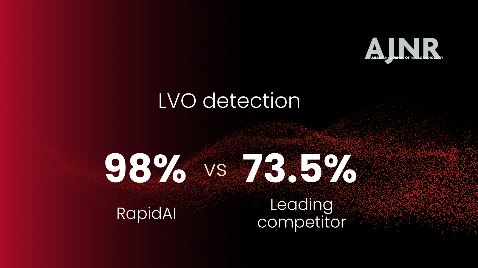


Dr. Sahlein and co-authors recently published a retrospective study using an AI-enabled aneurysm measurement tool, Rapid Surgical Preview, to calculate intracranial aneurysms' volume and surface area. Rapid Aneurysm had a higher sensitivity for detecting changes in aneurysm size than the current clinical practice or manual linear measurement.
The study findings have major implications for clinical practice, especially for conservatively managed aneurysm patients.
Dr. Daniel H. Sahlein, Dr. Alex Spiotta, and Dr. Greg Albers discussed the findings of the paper and the profound impact of Rapid Surgical Preview in a recent webinar.
Significance of the Rapid Surgical Preview volumetric tracking features
When treating an intracranial aneurysm, physicians weigh the risk and benefits between patient history and treatment.
Some of the factors considered to evaluate aneurysm rupture risk include smoking, a prior rupture from another aneurysm, family history, aneurysm growth, and irregular shape.
Though a change in aneurysm size and morphology are important risk factors, the current practice of using a single linear maximum dimension to measure the aneurysm size fails to capture the aneurysm size accurately. It doesn't consider the dynamic nature of aneurysms.
In addition, aneurysms often come in unusual morphologies, for which one linear dimension, or sometimes even multiple linear dimensions, really fails to estimate the size accurately. And smaller aneurysms can be challenging to measure.
Dr. Sahlein's publication showed how Rapid Surgical Preview tools could help physicians better assess rupture risk. The AI-enabled measurement tool detected changes in aneurysm size that were missed by manual linear measurement.
How does Rapid Surgical Preview work? From a patient's imaging scans, the platform creates a personalized 3D model of the vasculature and provides tools for accurate measurement and identification of morphological changes.
It calculates reproducible volumetric and surface area measurements, a more accurate reflection of aneurysm size and hence aneurysm growth.

Figure 1. Sagittal CTA maximum intensity projections from the initial exam (A) and 1 year later (B). Rapid Aneurysm outputs of the initial (C) and 1-year scans (D), side by side. (E) This panel shows the initial and 1-year scans overlaid, demonstrating aneurysm growth. Image source http://dx.doi.org/10.1136/jnis-2022-019339
The future of aneurysm management
Several questions persist when conservatively managing aneurysm patients. At what size should an aneurysm be treated? When are changes in size and morphology considered substantial enough to necessitate intervention?
According to Dr. Sahlein, even some of the most-cited aneurysm prospective and retrospective studies have failed to consider the dynamic nature of aneurysms. Some of the studies were carried out under the implicit assumption that aneurysm size does not change.
Rapid Surgical Preview has opened up a new way of modeling and studying aneurysms.
Dr. Sahlein and Dr. Spiotta believe that as large data sets are collected using tools like Rapid Surgical Preview, which more accurately reflects the aneurysm morphology, we can answer some of the questions that have challenged physicians when conservatively managing aneurysms.
Watch the RapidAI Research Series webinar and learn more about Rapid Surgical Preview, an AI-enabled, volumetric aneurysm measurement tool, and the results of our latest publication. Read how clinical leaders in the field are reacting to this ground-breaking study here.