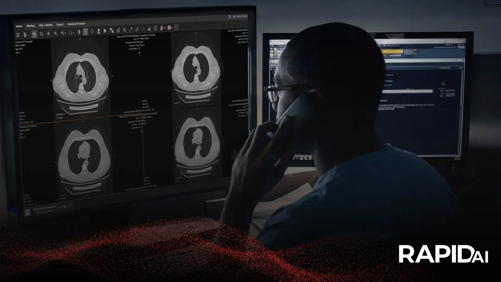

Every day, radiologists review thousands of chest imaging results ordered for pneumonia, pulmonary embolism, trauma, or unrelated post-operative follow-up. Buried in that anatomy, often unremarked upon in the referring clinician's original question, sits the aorta. It rarely asks for attention. Yet aortic pathology, when missed, can turn into one of the most catastrophic outcomes in emergency medicine.
Chest imaging was never designed with the aorta as its primary target in most of these studies. That is precisely why aortic disease so often slips past first review. Understanding how and why this happens is the first step toward closing the gap.
The aorta doesn't always announce itself via chest imaging
Thoracic aortic aneurysms are frequently silent. Most patients carrying one have no symptoms at all, which is exactly why these aneurysms tend to surface as incidental findings on chest imaging ordered for a completely different reason, whether a chest x-ray, a CT scan, or an echocardiogram performed for another clinical question.
That silence is part of what makes the aorta clinically dangerous. Without a triggering symptom, there is no prompt for a dedicated aortic workup. The diagnosis depends entirely on someone noticing it during a scan meant to answer a different question.
Why incidental findings are easy to miss
The scale of the challenge becomes clearer with the numbers. Aortic disease shows up incidentally on chest CT in as many as 3.4 percent of studies, and in one series, 21 of 22 incidentally detected aortic findings turned out to be aneurysms. In patients with atrial fibrillation undergoing contrast-enhanced chest CT, close to one in five scans showed a dilated aortic root or ascending aorta, with a subset large enough to meet aneurysm criteria.
These are not rare zebra cases. They are a recurring background presence in high-volume chest imaging, competing for attention with the primary indication for which the study was ordered. A radiologist working through a busy worklist, focused on ruling out pulmonary embolism or characterizing a lung nodule, is scanning past the aorta rather than scanning for it. Fatigue, volume, and divided attention all work against consistent detection of something that was never the stated reason for the exam.
The consequences of a missed or delayed diagnosis are significant. Aortic aneurysms tend to grow, and growth carries risk. Descending thoracic aneurysms above 6 centimeters carry a meaningfully elevated risk of rupture over time, and once symptoms appear, especially with dissection, the window for safe intervention narrows fast.
What the data reveals about detection gaps
Professional guidance has taken this seriously enough to formalize how radiologists should handle these findings. The American College of Radiology's Incidental Findings Committee published a white paper addressing exactly this scenario. Furthermore, committee members outlined specific criteria for measuring, classifying, and reporting incidental aortic dilation found on chest CT. Additionally, guidance calls for documenting anatomic location and precise diameter and distinguishing true aneurysmal disease from the softer categories of dilated or ectatic aorta that still warrant follow-up.
The existence of that guidance tells its own story. A detection and reporting framework this detailed does not get built for a rare event. It gets built because aortic pathology is common enough in everyday chest imaging that inconsistent reporting was becoming a real patient safety concern.
Bringing consistency to aortic detection in chest imaging
This is where purpose-built AI support changes the equation. Rather than relying solely on a radiologist catching an aortic finding while focused elsewhere, AI-driven aortic detection tools can now flag and measure aortic pathology in the background of routine chest imaging, surfacing dilation, aneurysm, or dissection findings for radiologist review regardless of what the specialists originally ordered to evaluate.
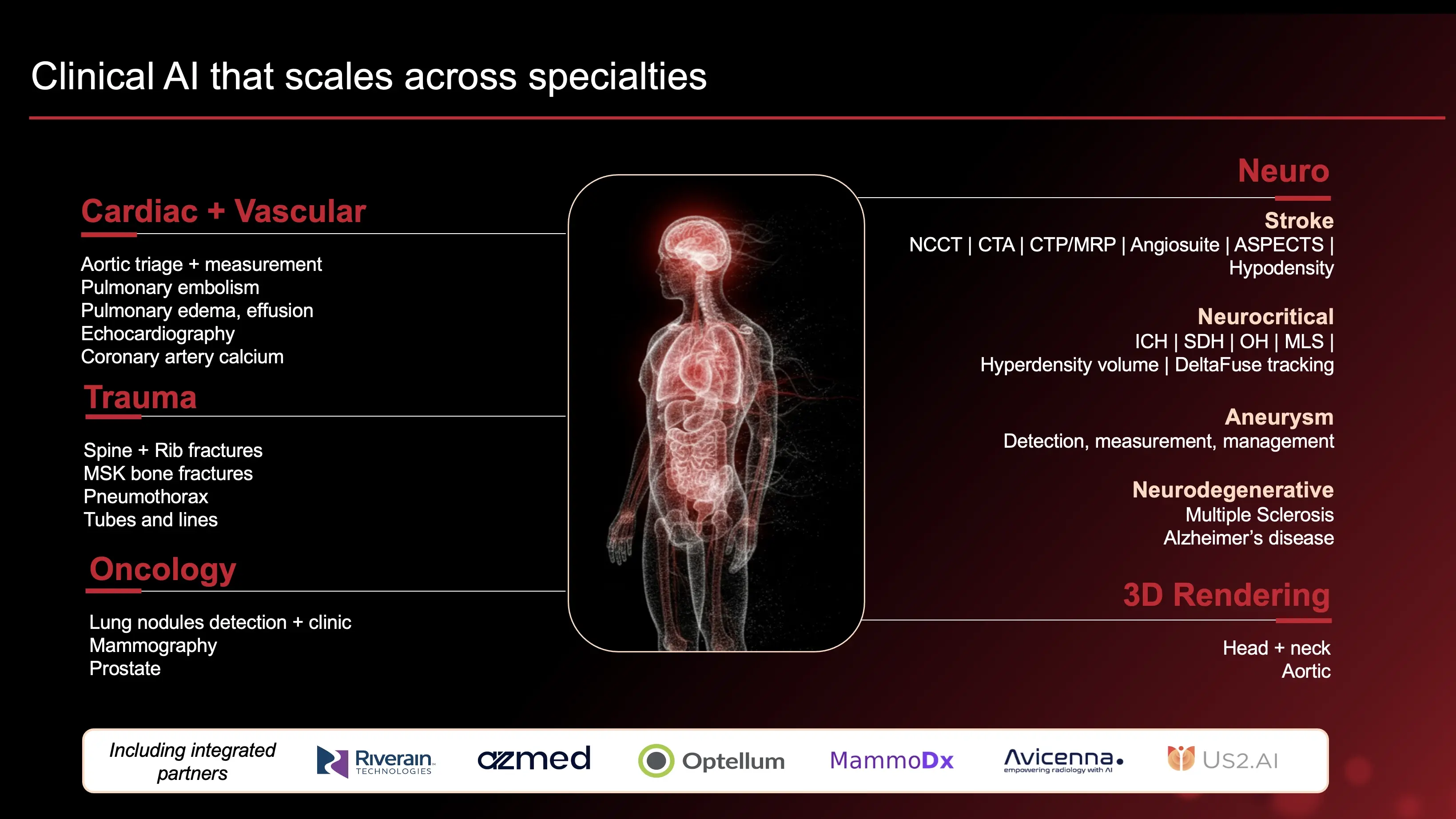
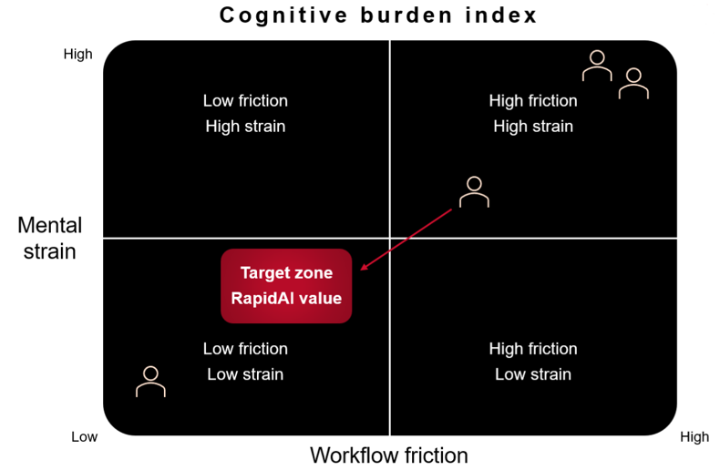
Radiology solutions built for high-volume workflows, such as tools that bring AI insights, measurements, and prioritization directly into the reading environment, help radiologists catch what competing demands on attention might otherwise let slip. And at the enterprise level, a scalable AI imaging infrastructure ensures consistency in aortic detection. This should not be limited to a single reader, shift, or site. It becomes a standard applied across every chest study, every day, at every location in a health system.
The same clinical rigor that has long defined stroke and vascular imaging AI is now extending into the aortic and cardiothoracic space. This gives radiology departments a way to close the detection gap that incidental findings guidelines should address.
A second look worth having
Aortic pathology hiding in routine chest imaging is not a hypothetical risk. It is a documented, measurable pattern with real consequences when missed, and rupture carries a mortality rate near 90 percent. Thankfully, RapidAI has an aortic solution built directly for this problem. It automatically analyzes every CT scan that includes the aorta, before or after treatment and works with both contrast and non-contrast scans. It generates guideline-based zonal and landmark measurements in moments and compares current and prior scans to identify changes over time, providing a comprehensive patient view.
If your department is looking for a more consistent way to catch these findings before they become emergencies, request a demo and see how our intelligent automation solutions could transform your workflow.
Frequently asked questions
Why is aortic disease often missed on routine chest imaging?
Most chest CT scans, x-rays, and echocardiograms are ordered for pneumonia, pulmonary embolism, trauma, or post-operative follow-up — not to evaluate the aorta. Thoracic aortic aneurysms are also frequently asymptomatic, so there's rarely a clinical trigger prompting a dedicated aortic workup. Radiologists focused on the primary indication, combined with high case volume and reading fatigue, can miss an aortic finding that was never the stated reason for the exam. Since detection depends entirely on incidental notice, aortic disease remains an easily overlooked background presence in routine chest imaging.
How common are incidental aortic aneurysms found on chest CT?
Incidental aortic findings are more common than many clinicians assume. Aortic disease appears incidentally on up to 3.4 percent of chest CT studies, and in one series, 21 of 22 such findings were true aneurysms. Among patients with atrial fibrillation undergoing contrast-enhanced chest CT, nearly one in five scans showed a dilated aortic root or ascending aorta. This is a consistent, measurable pattern rather than a rare event — and the stakes are high, since descending thoracic aneurysms above 6 centimeters carry meaningfully elevated rupture risk, and rupture itself carries a mortality rate near 90 percent.
How does AI help detect aortic aneurysms and dissections in chest imaging?
AI-powered aortic detection tools remove the reliance on a radiologist noticing an aortic abnormality while focused on an unrelated clinical question. Purpose-built AI can automatically analyze every CT scan that includes the aorta — contrast or non-contrast, before or after treatment — flagging dilation, aneurysm, or dissection for review regardless of why the scan was ordered. These tools generate guideline-based measurements in moments and compare current scans against prior imaging to track change over time. At the enterprise level, this brings consistent aortic detection to every chest study, every day, across a health system. RapidAI offers this kind of aortic solution, built to close that detection gap.