
Why ICAS-related LVO requires early recognition and better biomarkers
Intracranial atherosclerotic stenosis (ICAS) is a common cause of large vessel occlusion (LVO). However, accurately diagnosing ICAS-related LVO remains challenging in the acute stroke setting.
Endovascular thrombectomy (EVT) has become the standard of care for acute stroke patients with LVO. But, EVT is less efficacious in patients with underlying ICAS, making it essential to diagnose ICAS as quickly as possible before proceeding to treatment. Currently, there are no universally accepted biomarkers to predict ICAS with high accuracy.
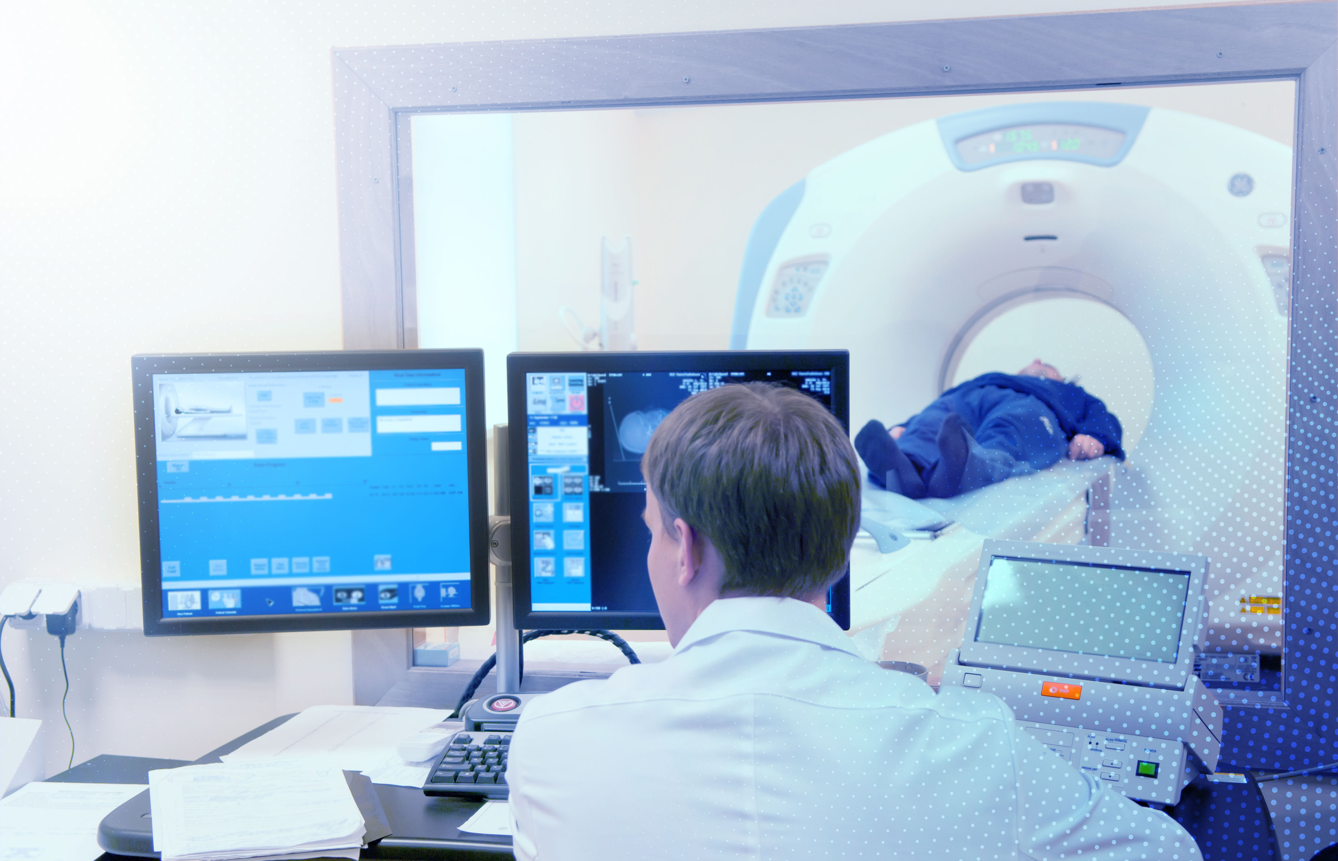
CT perfusion (CTP), which is now part of the modern stroke workflow, provides essential information on collateral status using biomarkers such as hypoperfusion intensity ratio (HIR) and cerebral blood volume (CBV) index. Patients with ICAS are known to have better collateral flow than those with other stroke subtypes.
This raises a critical question: Can HIR and CBV index provide clinicians with a reliable method to predict underlying ICAS before intervention?
A recent study led by Dr. Yukihiro Imaoka and co-authors hypothesized that lower HIR and higher CBV index would be helpful biomarkers to predict underlying ICAS before EVT.
Key takeaways:
- In an ROC analysis (receiver operating characteristic analysis), HIR showed good discrimination with a cutoff value of 0.22 (area under the curve, 0.85; 95%CI, 0.75−0.96; sensitivity, 0.84; specificity, 0.80) for underlying ICAS.
- CBV index showed excellent discrimination with a cutoff value of 0.90 (area under the curve, 0.92; 95%CI, 0.81−0.98; sensitivity, 0.92; specificity, 0.79) for underlying ICAS.
Study design and key findings on HIR and CBV index in ICAS-related LVO
This retrospective study was based on a single-site registry of consecutive LVO-acute ischemic stroke patients receiving EVT between October 2019 and December 2021.
Inclusion criteria:
- Patients with anterior circulation LVO (ICA, M1, and M2 proximal)
- Patients who received CTP imaging before EVT.
- CTP images were automatically analyzed using Rapid CTP.
Definitions:
HIR: Defined as the ratio of the volume of the Tmax >10 s lesion divided by the volume of the Tmax >6 s lesion.
CBV index: Defined as the ratio of the mean CBV within the "Tmax > 6 s lesion" in the ipsilateral hemisphere over the mean CBV of the ipsilateral area.
Patient breakdown:
Among the 238 patients who underwent thrombectomy in this period, 47 met the inclusion criteria. Out of the 47 patients, 10 patients (21%) had ICAS, and 37 (79%) had an embolism.
Findings:
Compared with the embolism group, patients with ICAS-related LVO demonstrated:
- A smaller ischemic core (0 ml vs. 18 ml; p = 0.002)
- Smaller Tmax > 10 s lesion (14 ml vs. 51 ml; p = 0.005)
- Lower HIR (0.12 vs. 0.50; p = 0.001)
- Higher CBV index (0.9 vs. 0.7; p < 0.001) than the embolism group
Diagnostic performance:
- In a receiver operating characteristic (ROC) curve analysis, HIR showed good discrimination with a cutoff value of 0.22 (area under the curve, 0.85; 95%CI, 0.75−0.96; sensitivity, 0.84; specificity, 0.80) for underlying ICAS.
- CBV index showed excellent discrimination with a cutoff value of 0.90 (area under the curve, 0.92; 95%CI, 0.81−0.98; sensitivity, 0.92; specificity, 0.79) for ICAS.
- Multivariable logistic regression analysis showed that an HIR ≤ 0.22 (OR, 22.5; 95%CI, 2.9 −177.0; p = 0.003) and a CBV index ≥ 0.9 (OR, 75.7; 95%CI, 5.8−994.0; p < 0.001) were significantly associated with underlying ICAS.
Conclusion:
These results suggest that an HIR ≤ 0.22 and a CBV index ≥ 0.9 may serve as reliable imaging biomarkers to predict ICAS-related LVO before thrombectomy.
The RapidAI Clinical Success team works closely with physicians and healthcare systems around the world to advance stroke care. Rapid CTP is the only FDA-cleared software to aid patient selection for acute stroke therapy.