Skip to content

Core Capabilities
-
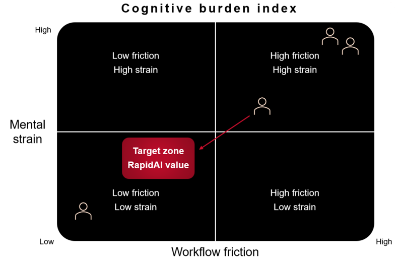
Deep clinical AIGoes beyond detection to surface deeper insights, + support more informed decisions
-
Workflow integrationIntegrates with EHR, PACS, and workflows to enable seamless clinical execution
-
Enterprise infrastructureScales securely to deliver high-performance clinical AI across the system
-
Data + analyticsProvides visibility into performance, utilization, and impact to optimize outcomes
Featured
Neurovascular
-
NeurocriticalFull suite of tools for neurocritical assessment, spanning ICH + hyperdensity, SDH, MLS, OH, and DeltaFuse
-
Ischemic strokeThe only complete solution across the patient journey, spanning NCCT, CTA, CTP, and intervention
-
AneurysmAI-driven detection support, growth assessment, and longitudinal tracking for rupture risk stratification
Featured
BUILT TO SUPPORT THE FULL SYSTEM
-
PhysiciansMove from imaging to action with decision-grade analysis, quantification, and clinical context
-
Care teamsAct faster with shared imaging insights, real-time collaboration, and coordinated care across teams
-
Hospital administratorsOperationalize AI with visibility into performance, utilization, and clinical impact across service lines
-
ITFits into your existing stack with secure, vendor-agnostic integration and scalable infrastructure with minimal lift
FEATURED
Core Capabilities
-
BlogClinical AI perspectives, product news, and healthcare technology insights
-
WebinarsLive and on-demand sessions with clinical experts and RapidAI leaders
-
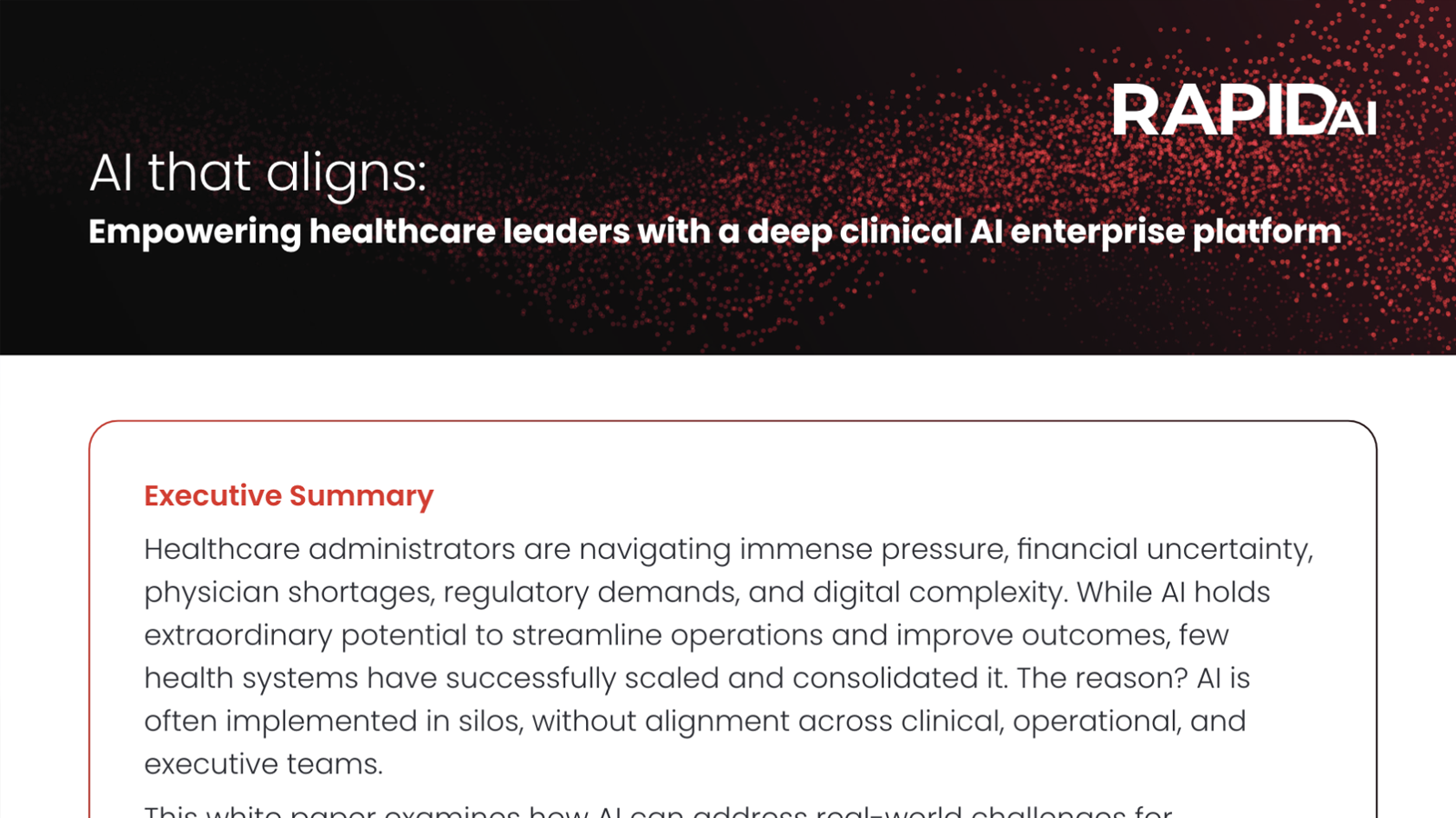
White papersDeep-dive on AI performance, evidence, and impact
-
VideosProduct demos, customer stories, and educational content
-
Inspiring outcomesReal stories of patient lives changed by faster, more connected care
-
Radiology Rewired podcastLeading clinicians, researchers, and industry disruptors unpack the factors that are redefining the future of imaging
Featured
LEARN MORE ABOUT RAPIDAI
-
Clinical validationThe research that laid the foundation for clinical AI across the enterprise
-
Publication library750+ peer-reviewed studies make RapidAI the most validated imaging AI platform
-
News + eventsCompany milestones, live + on-demand events, and conference presence
-
RapidAI blogAI in healthcare—insights, perspectives, and trends shaping the future of care
-
LeadershipThe team driving the future of AI-driven clinical decision support and care delivery